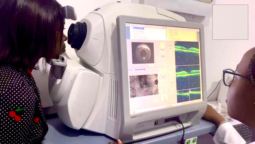
OCT, or Optical Coherence Tomography, is a non-invasive imaging technique used to visualize and analyze the microstructure of biological tissues, particularly the retina and optic nerve in the eye. OCT uses light waves to generate high-resolution, cross-sectional images of the retina, allowing for detailed assessment of retinal layers, thickness, and pathological changes. This imaging technology is invaluable in diagnosing and managing various eye conditions, including:
Macular Degeneration: OCT helps assess the integrity of the macula, the central part of the retina responsible for sharp, central vision. It can detect signs of age-related macular degeneration (AMD), including drusen deposits, retinal pigment epithelial changes, and fluid accumulation (edema) in the macula.
Diabetic Retinopathy: In diabetic retinopathy, OCT is used to detect and monitor changes in retinal thickness, microaneurysms, intraretinal hemorrhages, and macular edema caused by diabetes-related vascular damage. OCT imaging helps guide treatment decisions, such as intravitreal injections or laser therapy, to reduce macular swelling and prevent vision loss.
Glaucoma: OCT aids in the diagnosis and management of glaucoma by assessing the retinal nerve fiber layer (RNFL) and measuring optic nerve head parameters. Changes in RNFL thickness detected by OCT can indicate early glaucomatous damage and help monitor disease progression over time.
Retinal Edema and Fluid Accumulation: OCT is sensitive to detecting subtle changes in retinal thickness and fluid accumulation, such as cystoid macular edema (CME) or subretinal fluid, which may occur in various retinal disorders, including retinal vein occlusions and inflammatory conditions.
Macular Holes and Epiretinal Membranes: OCT imaging provides detailed visualization of structural abnormalities in the macula, such as macular holes and epiretinal membranes. It helps guide surgical planning and assess postoperative outcomes in patients undergoing vitrectomy for these conditions.
Retinal Detachment: OCT assists in diagnosing retinal detachment by visualizing the separation of retinal layers and detecting subretinal fluid accumulation. It helps ophthalmologists assess the extent of retinal detachment and determine the most appropriate surgical approach for repair.
Optic Nerve Disorders: OCT imaging of the optic nerve head aids in evaluating optic nerve head morphology, detecting changes in rim thickness, and assessing patterns of retinal nerve fiber layer loss in optic neuropathies such as optic neuritis, optic nerve drusen, or ischemic optic neuropathy.
Overall, OCT plays a critical role in the diagnosis, monitoring, and management of various eye conditions, enabling ophthalmologists to provide timely and targeted interventions to preserve vision and improve patient outcomes

