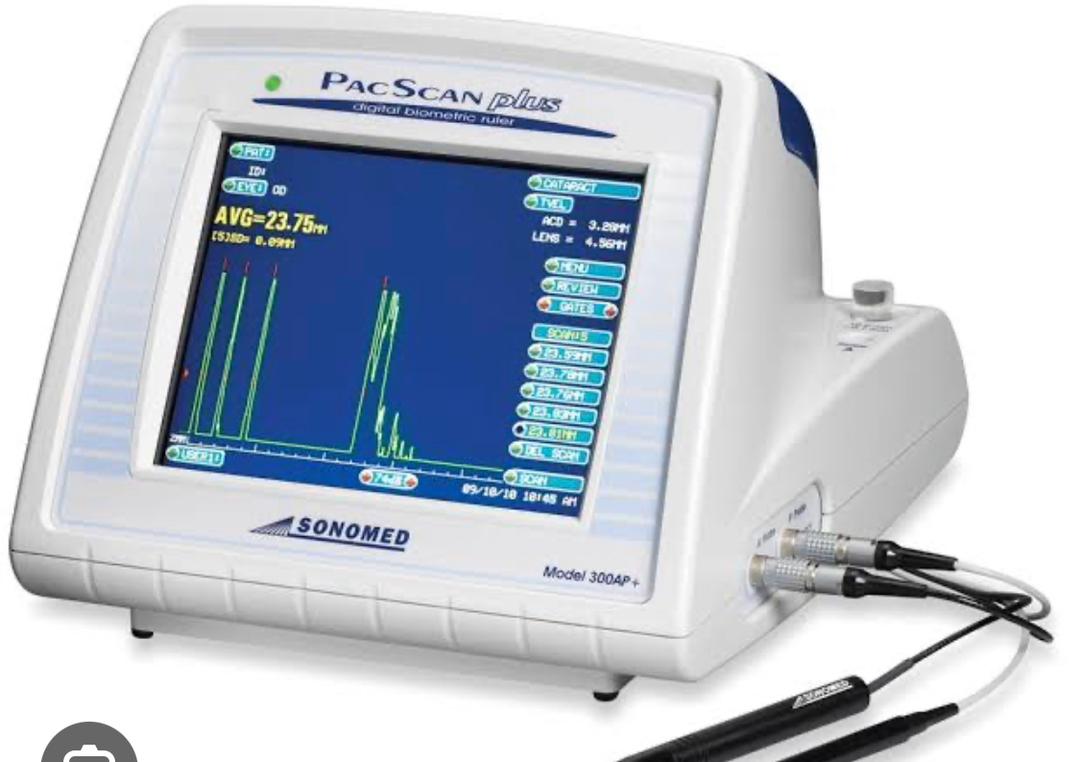
An A-scan ultrasound, or A-scan test, is a diagnostic procedure used to measure the length of the eye and assess the dimensions of ocular structures, particularly the anterior chamber depth and axial length. This non-invasive test provides valuable information for the evaluation of various eye conditions, such as cataracts, glaucoma, and refractive errors, and is commonly performed as part of preoperative assessment for cataract surgery and intraocular lens (IOL) calculations.
Purpose of A-Scan Test:
Cataract Evaluation: A-scan ultrasound helps assess the size and density of cataracts, which are clouding of the eye’s natural lens that can affect vision. By accurately measuring the axial length and thickness of the lens, A-scan provides essential information for determining the appropriate surgical approach and selecting the optimal power of the intraocular lens (IOL) to be implanted during cataract surgery.
Intraocular Lens (IOL) Calculations: In cataract surgery and certain refractive procedures, the natural lens of the eye is replaced with an artificial intraocular lens (IOL) to restore clear vision. A-scan measurements of axial length and anterior chamber depth are essential for calculating the power of the IOL needed to achieve the desired refractive outcome and minimize postoperative refractive errors such as myopia (nearsightedness) or hyperopia (farsightedness).
Glaucoma Assessment: A-scan ultrasound can be used to measure the anterior chamber depth and assess the angle structures in the eye, which are important parameters in the evaluation of glaucoma. Narrow angles or shallower anterior chambers may increase the risk of angle-closure glaucoma, a sight-threatening condition characterized by sudden increases in intraocular pressure.
Refractive Surgery Planning: Prior to undergoing refractive surgery procedures such as LASIK (laser-assisted in situ keratomileusis) or PRK (photorefractive keratectomy), patients undergo a comprehensive eye examination, including A-scan measurements, to assess ocular dimensions and suitability for surgery. Accurate measurement of axial length helps determine the amount of corneal tissue that can be safely removed during surgery to achieve the desired refractive correction.
Procedure for A-Scan Test:
During an A-scan ultrasound examination, the patient sits comfortably while a small probe, called a transducer, is placed gently on the surface of the eye after application of a lubricating gel. The transducer emits high-frequency sound waves that travel through the ocular tissues and are reflected back, creating echoes that are captured and analyzed by the ultrasound machine. Based on the time it takes for the sound waves to return and the speed of sound in the eye, the A-scan system calculates the dimensions of the eye structures, including axial length, anterior chamber depth, and lens thickness.
In summary, A-scan ultrasound is a valuable diagnostic tool in ophthalmology, providing essential measurements of ocular dimensions for the evaluation and management of various eye conditions, including cataracts, glaucoma, and refractive errors. By accurately assessing ocular parameters, A-scan testing helps optimize treatment outcomes and ensure the delivery of high-quality eye care.

